Researchers use mini lung models called organoids to explore how lung cancer starts

Carla Kim, PhD,
Boston Children’s Hospital Professor of Pediatrics in the Field of Regenerative Medicine
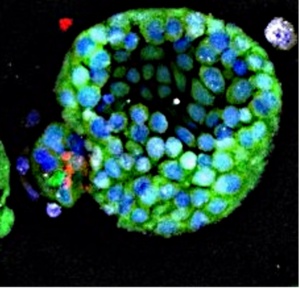
In the study “Organoid modeling reveals the tumorigenic potential of the alveolar progenitor cell state,” Li, Dang, Sengupta and colleagues used lung organoids, tiny lung-like structures grown in the laboratory dish from stem cells, to find new biomarkers and vulnerabilities of lung cancer in its earliest stages.
Starting with alveolar type 2 cells, the stem cells of the lung alveolar units that make breathing possible, the Kim Lab turned on the cancer-causing oncogene KRAS and used single cell sequencing to uncover which genes and proteins are activated just seven days after the oncogenic change.
The team discovered that even at the beginning of tumor formation, cancer cells are not identical–they are heterogeneous. Through their analysis, the researchers identified new biomarkers to identify the cells that drive cancer formation. By further analyzing the sequencing data, they uncovered several molecular pathways that are turned on in KRAS organoids, many of which have not yet been the focus of lung cancer therapeutic targeting. Notably, the study showed that blocking one of these factors, STAT3, can inhibit the growth of cancer organoids, suggesting that STAT3 may be a target in early-stage lung cancer.
The study highlights how studying the stem cells and the earliest steps in cancer formation can reveal new ways that drugs could target this deadly disease. Furthermore, organoid models are powerful “windows” into understanding how stem cells change to give rise to cancer, offering critical insights into lung cancer at its earliest stages—when interventions are most likely to stop tumor progression.


.